
[TargetMol Star Molecule] – PLX5622: A Precise Tool for Microglial Modulation in Neuroinflammation and Neurodegeneration Research
[TargetMol Star Molecule] – PLX5622: A Precise Tool for Microglial Modulation in Neuroinflammation and Neurodegeneration Research
■ 1. Background
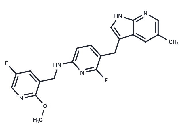
▲ PLX5622 (T7100),CAS: 1303420-67-8
PLX5622 (T7100), CAS: 1303420-67-8 is an orally active, brain-penetrant small-molecule inhibitor of the colony-stimulating factor 1 receptor (CSF1R; also known as c-Fms) that enables selective depletion of microglia in the brains of mice and rats. CSF1R is a receptor tyrosine kinase whose canonical ligands, CSF-1 and IL-34, drive receptor dimerization and autophosphorylation, initiating signaling through PI3K–AKT, ERK/MAPK, JAK/STAT, and Src-family kinase modules that collectively sustain myeloid-lineage survival, proliferation, metabolic fitness, motility, and transcriptional programs [1]. In the central nervous system, microglia express high levels of CSF1R and depend on tonic CSF-1/IL-34 signaling for viability, making this pathway a nodal control point for microglial maintenance [2].
By inhibiting the kinase activity of CSF1R/c-Fms, PLX5622 prevents receptor autophosphorylation and the recruitment/activation of downstream effector complexes. This interruption of pro-survival and proliferative cues collapses trophic support specific to microglia, leading to their efficient, brain-wide depletion in rodents. Functionally, the compound thereby modulates the c-Fms/CSF1R pathway upstream of key cytoprotective cascades (PI3K–AKT; ERK/MAPK) and transcriptional outputs (e.g., STAT-dependent programs), creating a controlled state of microglial absence. Oral bioactivity allows chronic or time-locked dosing paradigms that can be aligned with experimental triggers of neuroinflammation or injury, providing reproducible, selective ablation without surgical or genetic manipulation.
This pharmacological handle on CSF1R signaling has robust research utility. PLX5622 is widely used to establish microglia-deficient conditions for dissecting microglia’s causal roles in neuroinflammation, neurodegenerative disease models such as Alzheimer’s and Parkinson’s disease, traumatic or ischemic brain injury, and neuroimmune regulation. Depletion paradigms before or after an experimental insult help resolve stage-specific microglial contributions to inflammatory cascades, synapse remodeling, and neuron–glia crosstalk. Withdrawal of PLX5622 can be leveraged to study microglial repopulation and restoration of homeostatic networks, enabling analyses of resilience and plasticity in cellular and circuit phenotypes. Because CSF1R sits at the apex of lineage-defining signaling, modulation by PLX5622 allows researchers to uncouple microglial survival signaling from environmental cues and to parse microglia-dependent from microglia-independent mechanisms in complex CNS pathophysiology [1,2].
Operationally, PLX5622 integrates readily with histological, single-cell transcriptomic/epigenomic, and in vivo physiology readouts to quantify the consequences of microglial loss across scales—from cell-intrinsic stress responses and cytokine landscapes to network excitability and behavior. By targeting the c-Fms/CSF1R axis to extinguish the survival signals microglia uniquely require in the adult rodent brain, PLX5622 provides a precise and scalable approach to interrogate microglial biology and its system-level impact in models of neuroinflammation, neurodegeneration, brain injury, and neuroimmune regulation [2].
■ 2. Literature review
■ 2.1 Microglia Regulate Blood–Brain Barrier Integrity via MiR‐126a‐5p/MMP9 Axis during Inflammatory Demyelination
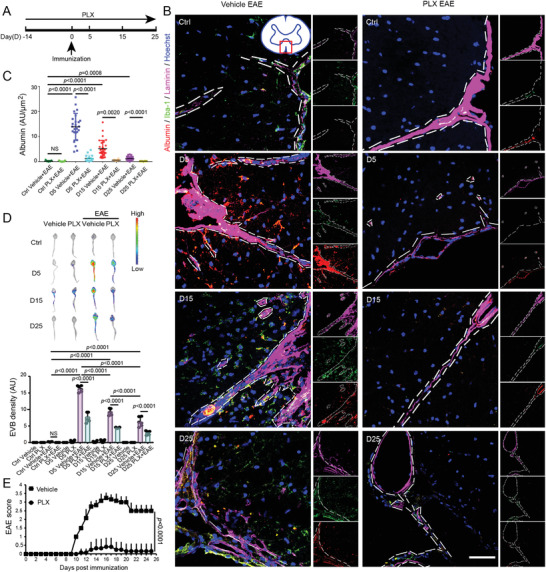
▲ Microglial ablation attenuates BBB impairment and ameliorates EAE progression. (A) Schematic of the timeline for microglial ablation, immunization, and examination. (B) Temporal dissemination of the BBB in the lumbar spinal cord of EAE mice following PLX5622 administration. Representative immunofluorescence images of Iba-1 (green), Albumin (red), Laminin (magenta, Alexa Fluor 647), and cell nuclei (blue) demonstrating the dynamic integrity of spinal cord coronal sections from EAE mice at early (Day 5 post immunization, D5), middle (D15), and late (D25) stages. Naïve mice served as controls (Ctrl); dashed lines indicate the boundary of Laminin+ vessels. Inset: spinal cord section with a box denoting the observed area. Scale bar: 50 µm. (C) Quantification of relative Albumin intensity in each group (Kruskal–Wallis test with Dunnett’s multiple comparisons test). (D) Representative optical imaging of BBB integrity in vehicle- or PLX5622-treated mice with or without EAE following i.v. Evans Blue (EB) injection (One-way ANOVA with Holm–Sidak’s multiple comparisons test). N=3 mice per group at each time point for immunofluorescence staining and EB extravasation assay. (E) EAE clinical score of mice administered PLX5622 or vehicle daily from 14 d before immunization to 25 d post immunization (Mann–Whitney test). N=9 mice per group for EAE score analysis. Data are shown as mean ± SEM.
PLX5622 (T7100) was used to eliminate microglia in an EAE mouse model, administered intragastrically every day starting 14 days before MOG immunization and continuing until sacrifice. After the last diphtheria toxin injection used for microglial ablation, PLX5622 was also administered to keep microglial depletion. In this setting, the EAE clinical score remained lower than in the control group when mice were treated with PLX5622 at 67.5 mg/kg, and even at 45 mg/kg. The study also examined timing by administering 90 mg/kg PLX5622 after the defined peak period of blood–brain barrier destruction on day 5 of EAE progression, and this later dosing was less effective to ameliorate EAE progression compared with PLX5622 given as pretreatment. The drug was prepared following the specified protocol, with stock solution dissolved in DMSO and diluted to a working solution prior to intragastric delivery, supporting consistent daily dosing throughout the experimental course.[3]
■ 2.2 Exosomes derived from M1-macrophage promote enteric neuronal injury via MMP8-TGF-β pathway
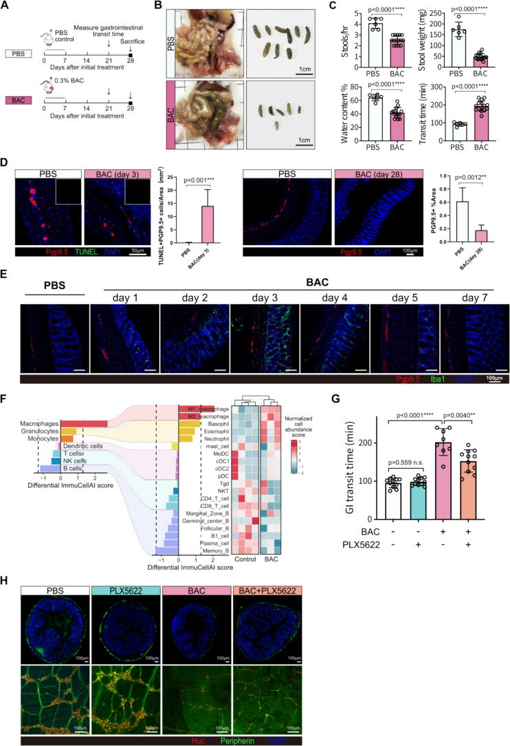
▲ BAC induces gastrointestinal dysmotility and macrophage infiltration in suckling mice, which was partially rescued by macrophage depletion. (A) Diagram illustrating the experimental design of the BAC mice model. (B) Representative images of the colons and feces of BAC-treated and control mice 4 weeks after treatment. (C) Quantitative results of the number of fecal pellets, feces weight, water content in fresh feces, and gastrointestinal transit time of the BAC and control mice. (D) Immunofluorescence (IF) analysis of neuronal apoptosis (TUNEL+ Pgp9.5+) in mice colons 3 days after treatment, and neuron density (Pgp9.5+) 4 weeks after treatment. (E) IF images showing the dynamic change of Iba1+ macrophages and Pgp9.5+ neurons after treatment. (F) Bar charts and heatmap representing relative immune cell abundance in mice colon samples treated with BAC or control estimated using the ImmuCellAI-mouse with RNA-seq data. (G–H) Macrophage depletion with PLX5622 partially reversed prolonged gastrointestinal transit time (G) and preserved the neuron density (Huc+, red) and plexus structure (Peripherin+, green) (H) in BAC mice.
PLX5622 (T7100) was used in this study as a selective CSF-1R inhibitor administered intraperitoneally to deplete macrophages in a benzalkonium chloride (BAC)-induced enteric neuronal injury mouse model. In this context, macrophage depletion with PLX5622 partially reversed the prolonged gastrointestinal transit time observed in BAC mice. In addition to the improvement in transit time, macrophage depletion with PLX5622 preserved neuron density and maintained the integrity of the enteric plexus structure, as indicated by Huc+ neuronal labeling and Peripherin+ network labeling. These findings were reported directly in BAC-treated mice subjected to macrophage depletion with PLX5622 and demonstrate that the compound, through targeted CSF-1R inhibition, was associated with improved gastrointestinal motility metrics and preservation of enteric neuronal architecture under injury conditions. The study explicitly describes the use of PLX5622 to achieve macrophage depletion and details the resulting beneficial outcomes on both functional (gastrointestinal transit time) and structural (neuron density and plexus structure) readouts in the injured gut.[4]
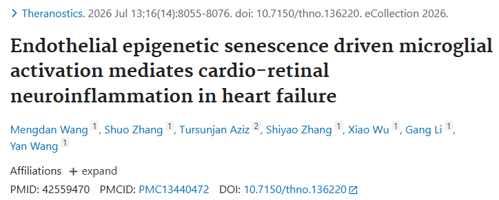
■ 2.3 Endothelial epigenetic senescence driven microglial activation mediates cardio-retinal neuroinflammation in heart failure.
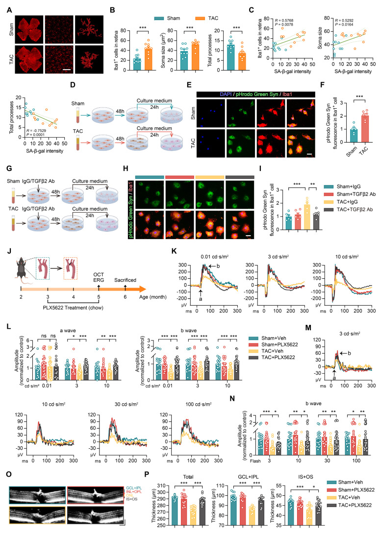
▲ Activation of microglia by senescent endothelial cells leads to retinal structural and functional damage, which is reversed by PLX5622-mediated microglial depletion. (A–B) Representative images and quantitative analysis of Iba1⁺ cell number, soma area, and total processes in the retinas of Sham and TAC mice (n = 10 mice per group; scale bar, 150 μm). (C) Correlation between Iba1⁺ cell number, soma area, total processes, and SA-β-gal intensity in retinal endothelial cells (n = 10 mice per group). (D) Schematic of the experimental timeline. (E–F) Representative images and quantitative analysis of pHrodo Green Syn fluorescence intensity in Iba1⁺ microglia after plasma treatment in Sham and TAC groups (n = 6 independent experiments per group; scale bar, 25 μm). (G) Schematic of the experimental timeline. (H–I) Representative images and quantitative analysis of pHrodo Green Syn fluorescence intensity in Iba1⁺ microglia and Iba1⁺ cell fluorescence intensity in different treatment groups (n = 6 independent experiments per group; scale bar, 25 μm). (J) Schematic of the experimental timeline: 2-month-old WT mice underwent Sham or TAC surgery, followed by PLX5622 treatment (1.2 g/kg, ad libitum) from 3 to 5 months. OCT and ERG were performed at 5 months, and tissue analysis was conducted at 6 months of age. (K–L) Scotopic ERG responses showing waveform plots and quantitative analysis of a-wave and b-wave amplitudes across groups (n = 20–40 eyes from 10–20 mice per group). (M–N) Photopic ERG responses showing waveform plots and quantitative analysis of b-wave amplitudes across groups (n = 20–40 eyes from 10–20 mice per group). (O–P) OCT cross-sectional images showing retinal structural changes with quantitative analysis (n = 14–20 mice per group; horizontal scale bar, 130 μm; vertical scale bar, 150 μm). All data are presented as mean ± SEM. P-values were determined using Student’s t-test in (B) and (F), one-way ANOVA with Dunnett’s post hoc analysis in (I) and (P), and one-way ANOVA with Tukey’s post hoc analysis in (L) and (N). ns, not significant; *p < 0.05, **p < 0.01, ***p < 0.001. The results demonstrate that PLX5622-mediated microglial depletion significantly ameliorates TAC-induced retinal functional deficits (e.g., restored ERG amplitudes) and structural damage (e.g., restored retinal thickness).
PLX5622 (T7100) was administered as a small molecule inhibitor of the colony-stimulating factor-1 receptor to eliminate microglia in Sham or TAC mice. Following administration, electroretinography demonstrated that PLX5622 improved ERG responses in TAC mice under dark-adapted testing. Under light-adapted conditions, PLX5622 increased b-wave amplitudes across different light intensities, indicating a protective effect on retinal function. Optical coherence tomography further showed that PLX5622 restored retinal layers that had been compromised in the disease context. Consistent with these functional and structural readouts, microglial depletion using PLX5622 interrupted the pathological process, leading to improved retinal integrity and function. Together, these findings show that PLX5622, through microglial elimination, confers measurable benefits on retinal function and structure in the experimental setting, enhancing ERG outcomes and reinstating retinal layer architecture while disrupting the underlying pathology and supporting retinal integrity.[5]
■ 3. Conclusion
PLX5622 functions by selectively inhibiting the kinase activity of CSF1R/c-Fms, thereby blocking receptor autophosphorylation and downstream pro-survival signaling cascades such as PI3K–AKT and ERK/MAPK. This leads to efficient, brain-wide depletion of microglia in rodents. Key findings from the literature demonstrate that PLX5622-mediated microglial depletion reduces disease severity in models of neuroinflammatory demyelination (EAE), preserves neuronal and plexus structure in enteric nervous system injury, and improves retinal function and architecture in cardio-retinal neuroinflammation. Moreover, timing and dosing of PLX5622 critically influence therapeutic outcomes, with earlier administration showing stronger benefits. Future applications may include refined temporal modulation of microglia to dissect stage-specific roles in CNS diseases, exploration of microglial repopulation dynamics post-treatment, and expanded use in diverse neuroimmune and neurodegenerative models for translational insights.
■ 4. Q&A
Q1: What is the mechanism by which PLX5622 depletes microglia in rodent brains?
A1: PLX5622 inhibits the kinase activity of CSF1R/c-Fms, preventing receptor autophosphorylation and downstream activation of pro-survival pathways such as PI3K–AKT and ERK/MAPK, leading to selective depletion of microglia.
Q2: How does the timing of PLX5622 administration affect its efficacy in EAE models?
A2: Administration of PLX5622 before the onset of EAE results in stronger amelioration of disease progression compared to dosing initiated after the peak of blood-brain barrier disruption.
Q3: Besides microglial depletion, what other cellular populations can PLX5622 target, and what are the associated functional outcomes?
A3: PLX5622 also depletes macrophages in models such as BAC-induced enteric neuronal injury, leading to improved gastrointestinal transit time and preservation of enteric neuronal architecture.
■ 5. Reference
[1] Stanley ER, Chitu V. CSF-1 receptor signaling in myeloid cells. Cold Spring Harb Perspect Biol. 2014;6(6):a021857.
[2] Elmore MRP, Najafi AR, Koike MA, Dagher NN, Spangenberg EE, Rice RA, et al. Colony-stimulating factor 1 receptor signaling is necessary for microglia viability, unmasking a microglia progenitor cell in the adult brain. Neuron. 2014;82(2):380-397.
[3] Yu Z, Fang X, Liu W, Sun R, Zhou J, Pu Y, et al.. Microglia Regulate Blood–Brain Barrier Integrity via MiR‐126a‐5p/MMP9 Axis during Inflammatory Demyelination. Advanced Science. 2022;9(24):.
[4] Chen Y, Song K, Zheng Y, Lan C, Mu Y, Ma Z, et al.. Exosomes derived from M1-macrophage promote enteric neuronal injury via MMP8-TGF-β pathway. Journal of Advanced Research. 2026;85():805-818.
[5] Wang M, Zhang S, Aziz T, Zhang S, Wu X, Li G, et al.. Endothelial epigenetic senescence driven microglial activation mediates cardio-retinal neuroinflammation in heart failure.. Theranostics. 2026;16(14):8055-8076.
Related products
-

Ordinary industrial grade polyethylene glycol
Contact UsPEG is a non-irritating, mildly bitter-tasting polyether compound with excellent water solubility and organic compatibility.
-
HZR818 Rutile TiO2 for Plastics & Masterbatch
Contact UsThis rutile titanium dioxide, produced via the sulfate process, is engineered to meet the demanding requirements of the plastics and masterbatch industries.
-
Cellin Vero Serum-free Medium Liquid
Contact UsProduct Name: Cellin Vero Serum-free Medium
Product Number: V6823C(Liquid) -

Sericin Powder
Contact UsFilament is the protein that constitutes the outer tissue of silk fiber, which constitutes silk together with the filin of the inner core.
Reviews
There are no reviews yet.